


|
Written by Michael Pettinati, PhD |
The Global Scope of Atrial Fibrillation
Atrial fibrillation (AFib) is a rapid, irregular atrial activation producing ventricular response and the most common sustained cardiac arrhythmia worldwide. Global prevalence nearly doubled between 1990 and 2019 to an estimated 59.7 million cases and is projected to rise further as populations age and risk factors such as hypertension, obesity, diabetes, and heart failure accumulate.1
In the United States, more than 10.5 million people live with AFib, a figure expected to reach 12.1 million by 2030.2 The condition drives more than 454,000 hospitalizations and contributes to roughly 158,000 deaths annually. AFib contributes to a fivefold increased risk of ischemic stroke and accounts for about one in seven strokes, which tend to be more disabling and more often fatal than those from other causes.2,3
For clinical development teams, these figures define both the opportunity and the challenge. AFib assessments are among the most complex in cardiovascular medicine, in large part because the disease is intermittent, heterogeneous, and frequently silent — while the conventional tools used to measure it were designed for an earlier era of cardiology.
The Detection Gap: Why Standard Tools Fall Short
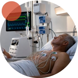 The current gold standard for detecting and measuring AFib is the 12-lead electrocardiogram (ECG), which provides a detailed 10-second snapshot of cardiac electrical activity from 12 different angles. This episodic assessment is generally adequate for persistent AFib, however in the case of paroxysmal AFib, which self-terminates and recurs unpredictably, it often misses episodes of AFib that occur in daily life. A routine ECG can show normal sinus rhythm in a patient carrying a clinically significant AFib burden — not because the arrhythmia has resolved, but because it was absent during those 10 seconds.
In a clinical study, this gap becomes endpoint unreliability. Rhythm outcomes assessed by scheduled clinic ECGs may reflect monitoring frequency as much as true disease status, producing systematic underestimation of AFib burden, reduced statistical power, and a structural bias toward null findings. Extended tools, such as 24-to-48-hour Holter monitors, 30-day event recorders, and implantable loop recorders, widen the observational window but remain episodic, burdensome to patients and sites, and, in the case of implantable devices, carry procedural risks that are impractical at digital trial scale.
Silent Episodes and the Limits of Symptom-Based Capture
AFib is a clinically variable condition. Some patients experience clear, debilitating symptoms, including palpitations, dyspnea, fatigue, and presyncope, that prompt clinical evaluation. However, many are entirely asymptomatic, with the arrhythmia discovered incidentally or only after a stroke. Others move between symptomatic and silent episodes, making self-reported diaries an unreliable surrogate for true burden. This variability is an important consideration for trial design. AFib burden is the proportion of monitored time in which AFib is detected and is widely regarded as the most clinically informative rhythm endpoint. Yet by definition, true AFib burden cannot be assessed using episodic monitoring tools. Because symptomatic patients are more likely to seek care, clinic-based monitoring is biased toward symptomatic disease and away from the asymptomatic burden that, in many populations, represents most of the total AFib time.
Where the Detection Gap Is Widest: Comorbidities
Heart Failure
Heart failure and AFib compound as reinforcing pairs, in which the presence of one worsens the prognosis of the other.14,15 AFib is the most common arrhythmia in heart failure, with prevalence ranging from 25% to 50% of patients depending on disease severity.14,15 Yet AFib in patients with heart failure is often paroxysmal and clinically silent against a chronic background of breathlessness and fatigue. Scheduled in-clinic ECGs only capture a fraction of the true rhythm picture, even though AFib onset, burden, and ventricular rate are among the variables that most directly drive poor outcomes. Because AFib is both highly prevalent and clinically consequential in heart failure, this population stands to benefit significantly from continuous monitoring, transforming what trials can measure beyond traditional episodic assessments.
Coronary Artery Disease
Coronary artery disease (CAD) and AFib share major risk factors and mechanisms and frequently coexist. AFib complicates a meaningful proportion of acute myocardial infarctions, while CAD raises the long-term risk of new-onset AFib through ischemia, atrial remodeling, and inflammation.16 When AFib appears in this setting, it independently worsens prognosis, raising the risk of recurrent ischemia, stroke, and death. The detection problem mirrors heart failure. When post-infarction patients are followed with continuous monitoring rather than scheduled ECGs, substantially more AFib is uncovered — and most of it is asymptomatic, precisely the burden that visit-based follow-up is structured to miss.
Obesity
Obesity is among the most important modifiable risk factors for AFib. In a large prospective cohort, obese individuals showed significantly elevated AFib risk relative to normal-weight controls, rising further with metabolic syndrome.4 Patients with obesity-related AFib are also more likely to have asymptomatic episodes, less likely to attend frequent in-clinic assessments, and more likely to show the reduced activity and elevated sedentary time independently linked to cardiovascular events and mortality.12
Post Surgery
Post-surgical AFib is among the most common complications of cardiac surgery, arising in roughly a third of patients, usually within the first days after an operation.17 Long treated as a transient, self-limiting nuisance, AFib is increasingly recognized as a marker of elevated stroke and mortality risk that frequently recurs after discharge, once monitoring has stopped. The SEARCH-AF trial made the gap concrete. Continuous rhythm monitoring in the 30 days after cardiac surgery detected A AFib in 19.6% of higher-risk patients versus 1.7% under usual care — an order-of-magnitude difference driven almost entirely by episodes that were paroxysmal, asymptomatic, and otherwise invisible.17 Because these patients are already instrumented and highly motivated, the post-operative window is a natural setting for passive, continuous wearable monitoring, and PPG-based rhythm surveillance after cardiac surgery is now being evaluated in randomized trials.18
Chronic Kidney Disease
Chronic kidney disease (CKD) and AFib share drivers including hypertension, diabetes, inflammation, and structural heart disease, and the presence of each accelerates the other. In the United States, age-adjusted mortality from comorbid AFib and CKD rose more than fourfold between 1999 and 2016.5 Anticoagulant therapy, the standard of care for stroke prevention, is especially complex in CKD. Impaired renal clearance alters drug pharmacokinetics, raising bleeding risk and complicating dosing. The result is a population in which the cost of AFib underdetection is highest, and conventional monitoring is least reliable.
Current AFib Detection and Management
The standard pathway for detecting and managing AFib begins with symptoms or an incidentally detected irregular pulse, followed by a 12-lead ECG, which is diagnostic only if the arrhythmia is active during recording. When the ECG is non-diagnostic, clinicians escalate to Holter monitoring (adequate for frequent symptomatic episodes, not for low-burden paroxysmal disease), extended event recorders (higher yield but dependent on adherence), or implantable loop recorders (highest yield but impractical for large-scale research).
| Strengths | Limitations | Best for | |
|
Holter Monitoring
|
|
|
Short term monitoring of patients with frequent or daily symptomatic episodes of an arrhythmia |
|
Implantable Loop Recorders (ILR) (up to 3 years)
|
|
|
High-risk patients with rare or unexplained events (i.e. cryptogenic strokes) after less invasive wearable options have been exhausted |
|
Event Recorders (14–30 day adhesive patches, mobile cardiac telemetry)
|
|
|
Weeks-long monitoring of infrequent symptoms and near real-time monitoring |
| Wrist-worn Wearable: PPG (Photoplethysmo-graphy) plus accelerometer |
|
|
Clinical trials and research studies requiring a long-term, scalable cardiac rhythm monitoring solution and screening tool |
Fit-for-Purpose AFib Monitoring with Photoplethysmography (PPG)
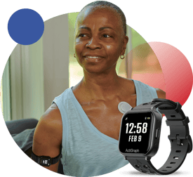 Photoplethysmography (PPG) is an optical method that measures blood-volume changes in peripheral tissue from differential light absorption through the skin. Wrist-worn watches, fitness trackers, and patch sensors run PPG continuously and passively, yielding pulse-to-pulse intervals — the wearable analog of the ECG R-R interval. In AFib, chaotic atrial activation produces characteristic pulse-interval variability that trained classifiers can identify without requiring P-wave resolution. Discriminating features include elevated Root Mean Square of Successive Differences (RMSSD) of interbeat intervals, increased sample entropy, and irregular pulse amplitude.
Early work by Lemay et al. (2016) showed that a wrist-worn infrared PPG device with a support-vector-machine classifier achieved 0.99 accuracy in a 20-patient study — comparable to pacemaker-based detection and well above implantable cardiac monitors (0.72).6 Fallet, Lemay et al. (2019) added wave-morphology and spectral features that reduced false positives from premature atrial and ventricular contractions, an advance for ambulatory settings where ectopy is common.7 Larger validations have since reinforced the performance:
-
-
Smartphone PPG reached 98.3% sensitivity and 99.9% specificity across real-world paroxysmal and persistent AFib.8
-
A 2024 smartwatch burden study (n=245; approximately 578,000 measurement intervals): 96.3% sensitivity, 99.5% specificity, and R²=0.996 against a Holter reference.9
-
A Cleveland Clinic, single-center 100-patient study revealed 94% sensitivity and 96% specificity, equivalent to single-lead ECG.10
-
The decisive operational advantage is that PPG offers continuous and passive AFib monitoring during daily life. Patients need not activate a device, recognize symptoms, or visit a clinical site to enable the detection of paroxysmal and asymptomatic AFib that episodic ECG misses by design. However, the method has limits. PPG is not as specific as ECG in diagnosing or characterizing arrhythmias because it is not measuring the heart’s electrical activity. Further, motion can introduce artifacts that require data exclusion. Best practice pairs PPG with accelerometry to exclude motion-contaminated segments and ECG for confirmatory rhythm characterization. Wearable PPG devices also allow for the derivation of additional signals, like HR, RR, and SpO2, in addition to AFib, which can provide insight into certain disease stages, acute adverse event onset, and patient deterioration. PPG-based monitoring affords certain signals that go beyond what ECG alone can provide.
A Regulatory Precedent: Apple Watch and the FDA Medical Device Development Tools (MDDT) Program
In May 2024, the Apple Watch atrial fibrillation history feature became the first digital health technology qualified under the U.S. Food and Drug Administration (FDA) Medical Device Development Tools (MDDT) program for a specific Context of Use as a secondary efficacy endpoint in cardiac ablation device trials.11 The feature uses PPG to estimate weekly AFib burden as a percentage of wear time. Qualification data showed 92.9% of subjects fell within 5 percentage points of an ECG-patch reference.11 MDDT qualification lets sponsors use a measurement tool as a trial endpoint without re-justifying its methodology from first principles in every protocol. The precedent establishes PPG-derived AFib burden as a regulatory-grade endpoint, and any sufficiently validated sensor-based measure now enters the conversation from that footing.
 However, the qualification does not erase the limitations sponsors must weigh. Consumer devices are subject to unilateral changes to firmware, algorithms, and hardware that can introduce measurement drift across multi-year, multi-site studies. They were not purpose-built for research, and PPG performance degrades in populations with darker skin tones, obesity, peripheral vascular disease, and high motion — all of which are often overrepresented in cardiovascular trials. Battery and charging requirements create monitoring gaps, wear adherence can be hard to enforce, and missing data may not be random. Cost, cross-border regulatory variation, and consumer data sharing add further complexity. With the endpoint category accepted and the validation framework defined, the open question for each protocol is whether a consumer device or a purpose-built research-grade monitor better serves its data-quality and regulatory requirements.
Actigraphy: Objective Functional Measures as Complementary Outcomes
Actigraphy — continuous accelerometer-based measurement of movement and activity — captures a dimension of cardiovascular health that rhythm monitoring does not. In the Prospective Urban Rural Epidemiology (PURE) study, physical activity and daily sedentary time were independent, dose-dependent predictors of cardiovascular events and all-cause mortality, robust to adjustment for traditional risk factors.12
For AFib research this matters for two reasons. First, AFib reduces cardiac output and exercise tolerance, so activity patterns can serve as sensitive functional indicators of burden and progression. Secondly, the comorbid populations where AFib is most complex, including heart failure, obesity, and CKD, are exactly those where reduced activity is most prevalent and most prognostic. Unlike activity questionnaires, which are subject to recall and social-desirability bias, actigraphy yields objective, time-stamped behavioral data collected in the patient’s own environment across the study assessment period.
Toward Integrated, Multimodal Monitoring
The value of combining PPG and actigraphy lies in their complementary data output. PPG characterizes how the heart is beating, while actigraphy captures how the patient is functioning. Together they offer a more complete and sensitive picture of cardiovascular health than either alone, and confirmatory single- or multi-lead ECG can be incorporated where clinical or regulatory requirements call for ECG-grade rhythm documentation. An emerging third signal is peripheral oxygen saturation (SpO₂). AFib with rapid ventricular response can impair cardiac output, and sleep apnea — bidirectionally linked to AFib — produces quantifiable nocturnal desaturation. Recent work validated wrist and upper-arm reflectance oximetry against polysomnography, reaching 1.9% root mean square error at the upper arm, within FDA and ISO thresholds for clinical-grade SpO₂ monitoring.13 This multimodal architecture is set to define the next generation of cardiovascular trials, with the Ametris science team currently working to validate signals across relevant patient cohorts.
Implications for Clinical Trial Design
Validated PPG performance, the FDA MDDT qualification of wearable AFib-burden measurement, and the established prognostic value of actigraphy together point to a clear framework for clinical development teams:
-
AFib burden as a primary or secondary endpoint — PPG-derived burden is now validated and regulatory-precedented for ablation outcomes trials, antiarrhythmic studies, and cardiovascular risk-reduction programs in comorbid populations.
-
Scalable patient monitoring in cardiovascular outcome trials — continuous monitoring enables systematic, prospective AFib detection where the arrhythmia is prevalent but undertreated, including heart failure, coronary artery disease, obesity, and other cardiovascular programs.
-
Decentralized, patient-centric design — passive, low-burden monitoring aligns with regulatory and payer interest in real-world evidence and reduced clinic burden, so sponsors no longer trade data quality for participant convenience.
Practical design considerations include pre-specified statistical analysis plans with defined clinically meaningful burden thresholds; device-validation documentation showing equivalence to the relevant MDDT-qualified reference; signal-quality and data-rejection criteria for PPG, particularly in high-motion and obese populations; and integration of actigraphy into pre-specified functional endpoints.
Conclusion
AFib is among the most significant unmet challenges in cardiovascular clinical research and development. It is prevalent, heterogeneous, and frequently silent, with standard monitoring that systematically underestimates true burden, particularly in the comorbid populations where the clinical consequences are greatest. Continuous wearable monitoring that combines PPG-based AFib burden detection, actigraphy monitoring, and emerging SpO₂ capability offers a digital measurement infrastructure suited to the disease. The technology is validated, the regulatory precedent is established, and the rationale for objective functional endpoints is well supported. The design question is no longer whether sensor-based monitoring can meet trial-grade standards, but how best to apply it to the population and endpoint at hand.
Explore Ametris' fit-for-purpose Cardiac Rhythm Monitoring Solution for AFib detection and cardiovascular research. Contact us to schedule a meeting with a member of our team to discuss your specific digital measurement needs.
References
1. Lippi G, Sanchis-Gomar F, Cervellin G. Global epidemiology of atrial fibrillation: an increasing epidemic and public health challenge. Int J Stroke. 2021;16(2):217–221. PMID: 31955707.
2. Centers for Disease Control and Prevention. Atrial Fibrillation Fact Sheet. CDC Division for Heart Disease and Stroke Prevention. 2024.
3. Wolf PA, Abbott RD, Kannel WB. Atrial fibrillation as an independent risk factor for stroke: the Framingham Study. Stroke. 1991;22(8):983–988. PMID: 1866765.
4. Wang TJ, Parise H, Levy D, et al. Obesity and the risk of new-onset atrial fibrillation. JAMA. 2004;292(20):2471–2477. PMID: 15479938.
5. Odutayo A, et al. Atrial fibrillation and risks of cardiovascular disease, renal disease, and death: systematic review and meta-analysis. BMJ. 2016;354:i4482. PMID: 27599725.
6. Lemay M, Fallet S, Renevey P, Leupi C, Pruvot E, Vesin JM. Bertschi M, Sola J, Renevey P, Parak J, Korhonen I. Wrist-located optical device for atrial fibrillation screening: a clinical study on twenty patients. Computing in Cardiology (CinC). 2016.
7. Lemay M, Fallet S, Renevey P, et al. Can one detect atrial fibrillation using a wrist-type photoplethysmographic device? Med Biol Eng Comput. 2019;57(12):2521–2532. PMID: 31643033.
8. Gruwez H, Ezzat D, Van Puyvelde T, et al. Real-world validation of smartphone-based photoplethysmography for rate and rhythm monitoring in atrial fibrillation. EP Europace. 2024;26(4):euae065. PMID: 38630867.
9. Perez MV, MahAFibfey KW, Hedlin H, et al. Large-scale assessment of a smartwatch to identify atrial fibrillation. N Engl J Med. 2019;381(20):1909–1917 (algorithm validation context). PMID: 31722151.
10. Bumgarner JM, Lambert CT, Hussein AA, et al. Smartwatch algorithm for automated detection of atrial fibrillation. J Am Coll Cardiol. 2018;71(21):2381–2388. PMID: 29535065.
11. U.S. Food and Drug Administration. FDA qualifies Apple Watch atrial fibrillation history feature under the Medical Device Development Tools program. FDA Digital Health Center of Excellence. 2024.
12. Lear SA, Hu W, Rangarajan S, et al. (PURE Study Investigators). The effect of physical activity on mortality and cardiovascular disease in 130,000 people from 17 countries: the PURE study. Lancet. 2017;390(10113):2643–2654. PMID: 29031021.
13. Lemay M, Braun F, et al. Continuous SpO₂ monitoring at wrist and upper arm during overnight sleep apnea recordings. IEEE Engineering in Medicine and Biology Conference (EMBC). 2025. PMID: 41336868.
14. Zuin M, Bertini M, Vitali F, Turakhia M, Boriani G. Heart failure-related death in subjects with atrial fibrillation in the United States, 1999 to 2020. J Am Heart Assoc. 2024;13(9):e033897. PMID: 38686875.
15. Karnik AA, Gopal DM, Ko D, Benjamin EJ, Helm RH. Epidemiology of atrial fibrillation and heart failure: a growing and important problem. Heart Fail Clin. 2019;15(4):447–455. PMID: 30926013.
16. Börschel CS, Schnabel RB. The imminent epidemic of atrial fibrillation and its concomitant diseases — myocardial infarction and heart failure. Int J Cardiol. 2019;287:162–173. PMID: 30528622
17. Ha ACT, Verma S, Mazer CD, et al.; SEARCH-AFIB CardioLink-1 Investigators. JAMA Netw Open. 2021;4(8):e2121867. PMID: 34448866.
18. Gruwez H, De Melio N, Vermunicht P, et al. Smartphone-based photoplethysmography rhythm monitoring following cardiac surgery: a pragmatic randomized trial. EP Europace. 2025;27(2):euAFib015.